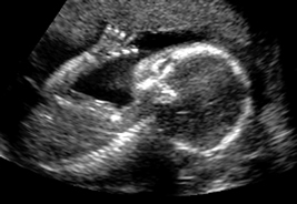
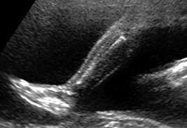
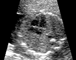
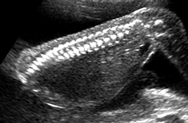
During your ultrasound, a picture of your baby is produced when high-frequency sound waves bounce off your baby and translate into an image on screen. In this image, solid matter, such as bones, are white, while softer tissue appears gray. Areas that contain fluid, such as blood vessels or the stomach, as well as the amniotic fluid, do not respond to the sound waves and therefore appear as black areas on the scan. The sonographer will study these details to assess how your baby is developing in the uterus.
Images of a 20-Week Ultrasound Scan
Join the Family
Your partner in parenting from baby name inspiration to college planning.
Subscribe


